Joint Effort Launched to Create AI-Powered Algorithm to Detect Hemorrhages in Brain Scans
CGI, Helsinki University Hospital, and Planmeca announced the partnership.


Artificial intelligence is once again being paired with imaging technology in the medical field.
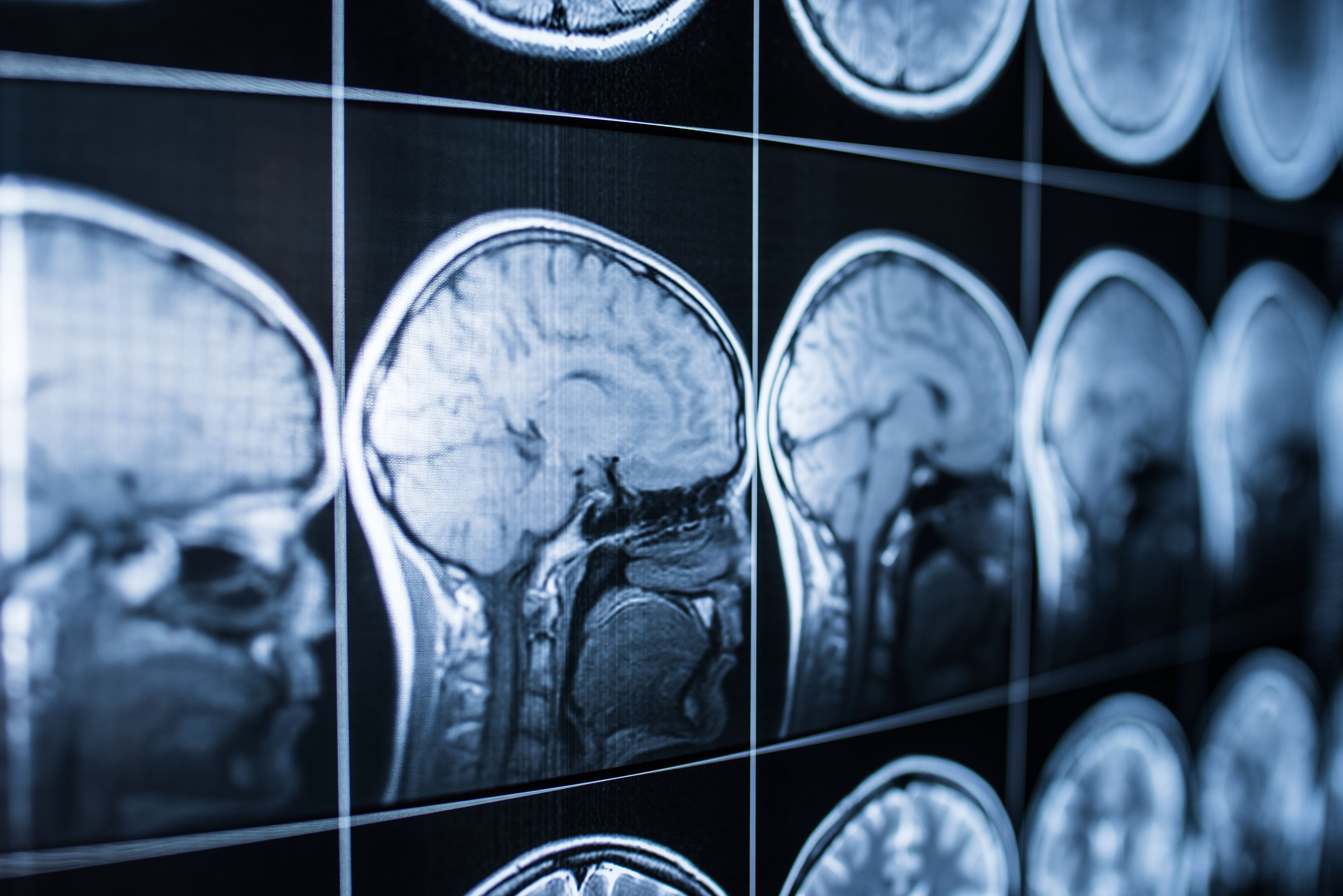
Three organizations (CGI, Helsinki University Hospital, and Planmeca) have teamed up to develop an imaging algorithm to be used with CT scans. The AI program will reportedly be capable of detecting many common types of non-traumatic brain hemorrhages.
In a joint press release, the organizations announced that the platform will be named AI Head Analysis and is part of CGI’s Clever Health Network. The algorithm is being designed to be used alongside radiologists, who will be able to use the platform to compare their results and diagnosis. It will pull data from multiple imaging devices to build its algorithms and has the potential to speed up the process of getting patients to the treatment stage.
In the press release, Miikka Korja, Chief Innovation Officer and a docent in neurology at the Helsinki University Hospital, said, “Each year, millions of people across the globe are diagnosed with some type of brain hemorrhage—an acute condition that requires a prompt and accurate diagnosis. In Finland alone, more than 180,000 head CT scans are performed annually, most of them at emergency clinics.”
Continue reading the press release here.
This is the latest example of an AI algorithm being used with imaging devices in the medical field. Medical Device & Technology recently spoke with Victor Chan, VP of marketing for Body Vision Medical, about his company’s algorithm designed to be used with C-arms.
These devices work similarly to CT scanners, although they take less images and have to be manually moved, meaning that they don’t take X-rays at the same pre-determined points as a CT scanner would. Body Vision Medical’s platform, however, can take these images and create a 3d model of the patient’s body. This provides doctors who don’t have access to larger CT scanners with higher quality images when making their diagnosis.
This program was recently used alongside Serpex’s steerable needle to successfully perform a lung nodule biopsy at Cleveland Clinic in Ohio.